Obesity Hypoventilation on Polysomnography
Practical notes for the Polysomnographic Technician / Sleep Technician preparing for the RPSGT exam. Learn to separate obesity-associated sleep hypoventilation from established obesity hypoventilation syndrome, follow oxygen and carbon dioxide across the night, recognize REM worsening, and connect PSG findings with treatment and technologist scope.
1. What Is Obesity Hypoventilation Syndrome?
For board-prep purposes, the standard definition combines obesity, sleep-disordered breathing, awake daytime hypercapnia, and exclusion of another cause of alveolar hypoventilation.
| Finding | Board-prep meaning |
|---|---|
| BMI ≥30 kg/m² | Meets the obesity component, but obesity alone does not diagnose OHS. |
| Sleep-disordered breathing | Most patients have OSA, although a smaller group has sleep hypoventilation without severe obstruction. |
| Awake PaCO₂ ≥45 mm Hg | Supports the daytime hypercapnia component required by the standard definition. |
| Other causes excluded | Consider lung disease, neuromuscular disease, chest-wall disorders, medications, metabolic causes, and other conditions. |
Interactive check: Does severe nocturnal disease automatically prove OHS?
A patient has a BMI of 43 kg/m², AHI 157 events/h, profound desaturation, and tcCO₂ above 50 mm Hg for 41 minutes. Awake CO₂ is normal.
2. Recognizing the Pattern on PSG and the Hypnogram
Airflow
Breathing may continue, but the breaths remain shallow and provide insufficient alveolar ventilation.
Effort
Thoracic and abdominal excursion may be reduced rather than showing repeated complete obstructive events.
SpO₂
Oxygen may drift downward gradually and remain low for prolonged periods.
CO₂
Transcutaneous or end-tidal CO₂ may rise progressively across sleep and worsen during REM.
Hypnogram
Look for repeated worsening during REM periods and a whole-night trend that discrete events do not fully explain.
Coexisting OSA
Apneas, hypopneas, snoring, and flow limitation can coexist with sustained hypoventilation.
3. Downloadable Two-Minute REM Study Sheet
AI-rendered educational simulation
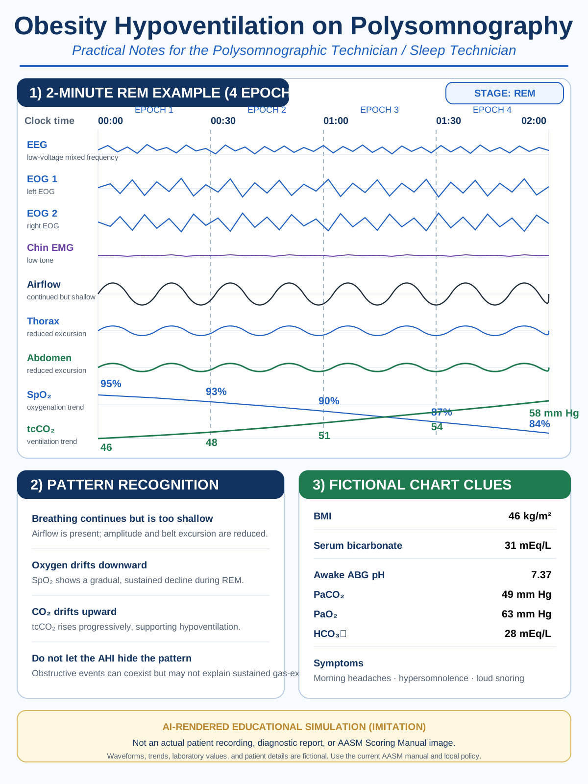
This original illustration imitates a two-minute PSG display with four 30-second REM epochs, two EOG channels, shallow airflow, reduced thoracoabdominal excursion, falling SpO₂, rising tcCO₂, and fictional chart clues.
 Download PNGDownload printable SVG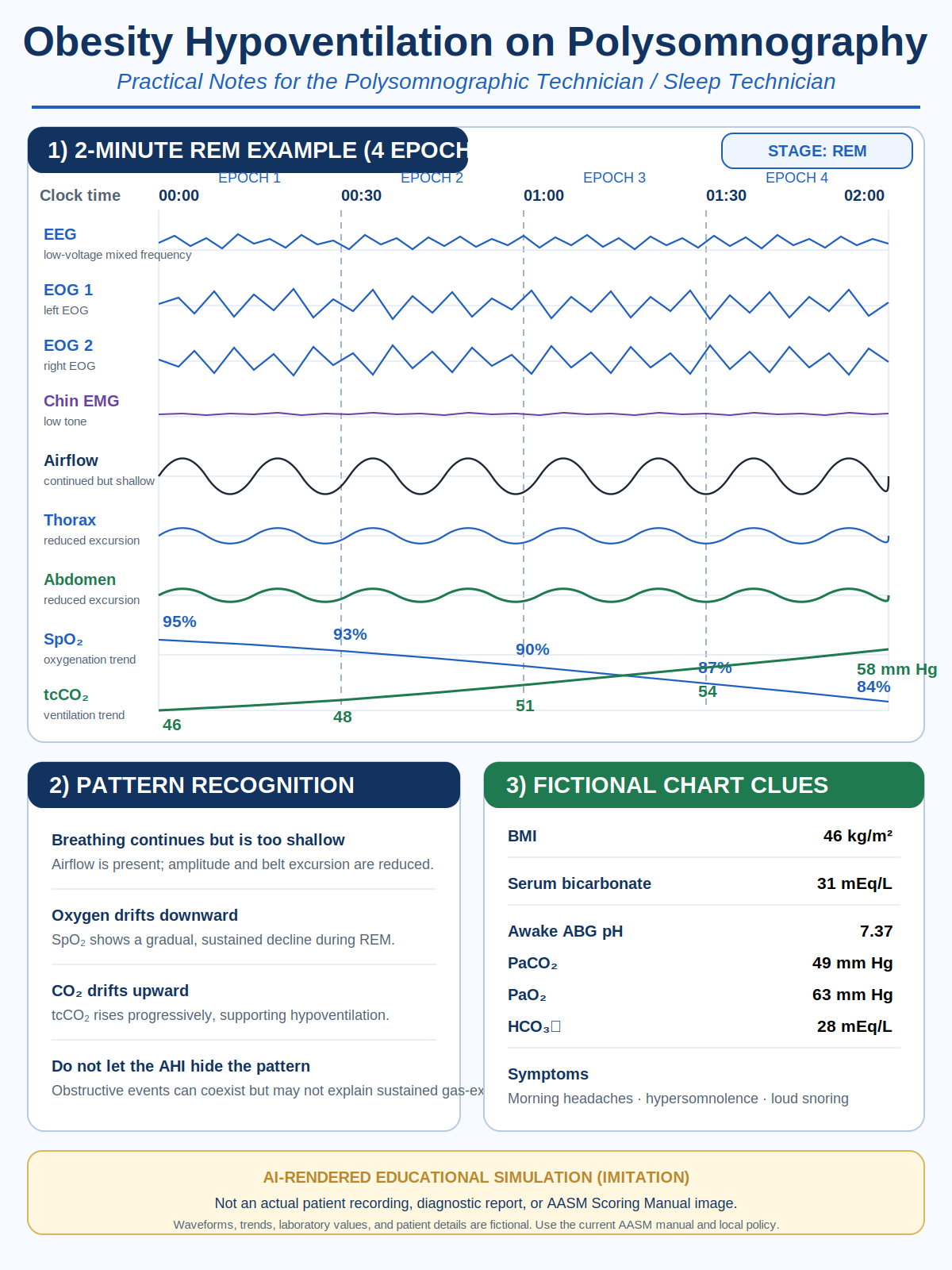
Image disclosure: AI-rendered educational simulation (imitation)—not an actual patient recording, diagnostic report, or AASM Scoring Manual image. Waveforms, values, and patient details are fictional.
4. Adult Sleep-Hypoventilation Scoring Anchors
For adult board-prep questions, commonly cited scoring anchors are:
- PCO₂ or an accepted surrogate above 55 mm Hg for at least 10 minutes, or
- an increase of at least 10 mm Hg from the awake supine value to a value above 50 mm Hg for at least 10 minutes.
Scoring drill 1
Awake supine tcCO₂ is 41 mm Hg. During sleep it rises to 52 mm Hg and remains there for 14 minutes. Does the pattern meet a commonly cited adult criterion?
Scoring drill 2
tcCO₂ remains at 56–58 mm Hg for 12 minutes. Awake baseline is unavailable.
Scoring caution: SpO₂ without CO₂
SpO₂ remains 84%–87% for six minutes in REM, but no CO₂ channel is recorded.
5. Chart and Blood-Gas Clues
| Chart clue | How to use it | Exam trap |
|---|---|---|
| Serum bicarbonate <27 mEq/L | Can make OHS less likely when suspicion is low to moderate. | Do not use it as a universal exclusion test in a highly suspicious patient. |
| Serum bicarbonate ≥27 mEq/L | Raises concern for chronic CO₂ retention and supports confirmatory awake gas evaluation. | Bicarbonate alone does not diagnose OHS and can be elevated for other reasons. |
| Awake ABG PaCO₂ ≥45 mm Hg | Supports the daytime hypercapnia component. | An overnight tcCO₂ value is not identical to an awake arterial PaCO₂. |
| Morning headache, hypersomnolence, loud snoring | Increase clinical suspicion when combined with obesity and abnormal gas exchange. | Symptoms are supportive, not diagnostic by themselves. |
6. Treatment Options the Sleep Technician Should Understand
CPAP
CPAP is commonly considered first when stable ambulatory OHS coexists with severe OSA. It stabilizes the upper airway and may improve gas exchange by preventing repeated obstruction. During titration, do not declare success from AHI control alone; also review breathing depth, CO₂, SpO₂, leak, stage, position, and patient tolerance.
Bilevel PAP and noninvasive ventilation
Bilevel support adds pressure support between IPAP and EPAP to assist tidal volume and minute ventilation. NIV is more likely when hypoventilation dominates, OSA is absent or mild, CPAP does not adequately correct gas exchange, or the patient has acute-on-chronic hypercapnic respiratory failure. Backup-rate and volume-assured modes are clinician- and protocol-directed options for selected patients.
Adding oxygen to PAP
Oxygen treats hypoxemia, not the underlying inadequate ventilation. The practical sequence is: verify signals, control obstruction, optimize ventilation, then add prescribed oxygen when clinically important hypoxemia persists. Continue monitoring CO₂ because a better SpO₂ number does not prove that ventilation has normalized.
Weight-management medication
Anti-obesity medications can support meaningful weight loss and may reduce the respiratory load that contributes to OSA and OHS. They do not replace PAP or NIV for established hypoventilation. Tirzepatide has an FDA indication for moderate-to-severe OSA in adults with obesity, but that is not the same as an OHS indication.
Surgery
Metabolic or bariatric surgery is the most relevant surgical strategy because sustained major weight loss can improve or resolve OHS. Upper-airway surgery may improve selected obstructive anatomy but does not automatically correct obesity-related mechanics, respiratory drive, or daytime hypercapnia. Tracheostomy is a rare rescue option rather than routine OHS therapy.
7. Original RPSGT-Style Practice Questions
These are original educational questions—not recalled or official BRPT examination items.
1. Which finding completes the standard OHS definition in an obese patient with sleep-disordered breathing?
- SpO₂ nadir below 80%
- Awake PaCO₂ at or above 45 mm Hg after excluding another cause
- AHI above 30 events/h
- Serum bicarbonate above 27 mEq/L
2. Which PSG pattern most strongly suggests sustained sleep hypoventilation?
- One abrupt desaturation after a position change
- Continued shallow breathing with rising CO₂ and gradual oxygen decline
- Repeated airflow cessation with continued effort only
- An isolated central apnea after an arousal
3. Why may the pattern worsen during REM?
- Respiratory effort always disappears
- Accessory-muscle activity is reduced and ventilation depends more heavily on the diaphragm and respiratory drive
- Oxygen consumption stops
- REM rules out OHS
4. The AHI is controlled on CPAP, but tcCO₂ continues rising and SpO₂ remains low in supine REM. What is the best first technologist action?
- Declare the titration successful
- Add oxygen without checking signals
- Verify airflow, effort, CO₂, SpO₂, leak, stage, position, and patient status, then follow the ordered protocol
- Diagnose respiratory failure
5. Why is oxygen alone inadequate treatment for OHS?
- It cannot raise SpO₂
- It may improve oxygenation without correcting inadequate ventilation or CO₂ retention
- It always worsens OSA
- It eliminates apneas
6. Which note is most appropriate?
- “Patient diagnosed with OHS.”
- “Patient needs home bilevel and oxygen.”
- “During supine REM, shallow breathing was accompanied by reduced belt excursion, rising tcCO₂, and sustained desaturation; signal quality was verified and ordered protocol followed.”
- “Pulmonary hypertension caused the events.”
8. Published Examples and Free Teaching Resources
Open OHS review
Includes an overnight example with persistent hypoxemia and worsening at likely REM periods.
Open reviewObesity-associated sleep hypoventilation with severe OSA
Whole-night summary with hypnogram, respiratory events, SpO₂, heart rate, and TCO₂.
Open case reportObesity and sleep-related hypoventilation
Supplementary material includes example hypnograms and five-minute PSG epochs.
Open studyATS OHS guideline resources
Guideline, summary, webinar, podcast, patient material, and clinician video.
Open ATS toolsNox Academy sleep-scoring series
Free scoring education; verify current recording access and any credit eligibility.
Open Nox AcademyCurrent AASM Scoring Manual page
Use the current manual and errata for final scoring language and technical specifications.
Open AASM scoring resources9. References
- Berry, R. B., et al. (2012). Rules for scoring respiratory events in sleep: Update of the 2007 AASM Manual. Journal of Clinical Sleep Medicine, 8(5), 597–619.
- Lin, J.-I. (2025). Obesity-associated sleep hypoventilation and concomitant severe obstructive sleep apnea. Journal of Sleep Medicine, 22(1), 32–34.
- Masa, J. F., et al. (2019). Obesity hypoventilation syndrome. European Respiratory Review, 28, 180097.
- Mokhlesi, B., et al. (2019). Evaluation and management of obesity hypoventilation syndrome: An official ATS clinical practice guideline. American Journal of Respiratory and Critical Care Medicine, 200(3), e6–e24.
- Piper, A. (2010). Obesity hypoventilation syndrome: Therapeutic implications for treatment. Expert Review of Respiratory Medicine, 4(1), 57–70.
- American Academy of Sleep Medicine. The AASM Manual for the Scoring of Sleep and Associated Events. Consult the current online manual and errata.