Learning goals
Identify a sustained hypoventilation pattern across PSG channels and whole-night trends.
Apply the adult CO₂ duration pathways using a valid awake supine baseline and a reliable surrogate.
Separate upper-airway obstruction, inadequate ventilation, and persistent hypoxemia.
Describe objective findings and follow the physician order, laboratory protocol, and technologist scope.
1. OHS is not diagnosed from the PSG alone
For board-prep purposes, obesity hypoventilation syndrome combines four elements:
BMI ≥30 kg/m².
Usually OSA, although sustained sleep hypoventilation may be the dominant pattern.
Awake resting PaCO₂ ≥45 mm Hg at sea level.
Other pulmonary, neuromuscular, chest-wall, metabolic, medication-related, or central causes must be evaluated.
2. What the polysomnogram may show
Hypoventilation is a ventilation problem over time. Breathing may continue, but the breaths may be too shallow to remove enough CO₂.
| Pattern | Airflow and effort | Gas-exchange clue |
|---|---|---|
| Obstructive apnea | Airflow stops or nearly stops while effort continues. | Discrete event with possible desaturation or arousal. |
| Obstructive hypopnea | Partial airflow reduction with continued effort. | Discrete event scored under the selected adult rule. |
| Sleep hypoventilation | Airflow and effort continue but may remain shallow or reduced. | CO₂ rises over minutes, often with sustained or progressive hypoxemia. |
- Review airflow, thorax, abdomen, SpO₂, tcCO₂ or EtCO₂, stage, position, leak, and patient appearance together.
- REM may reveal worsening because accessory respiratory muscle activity is reduced.
- OSA and sustained hypoventilation can coexist; the AHI may not explain the entire gas-exchange abnormality.
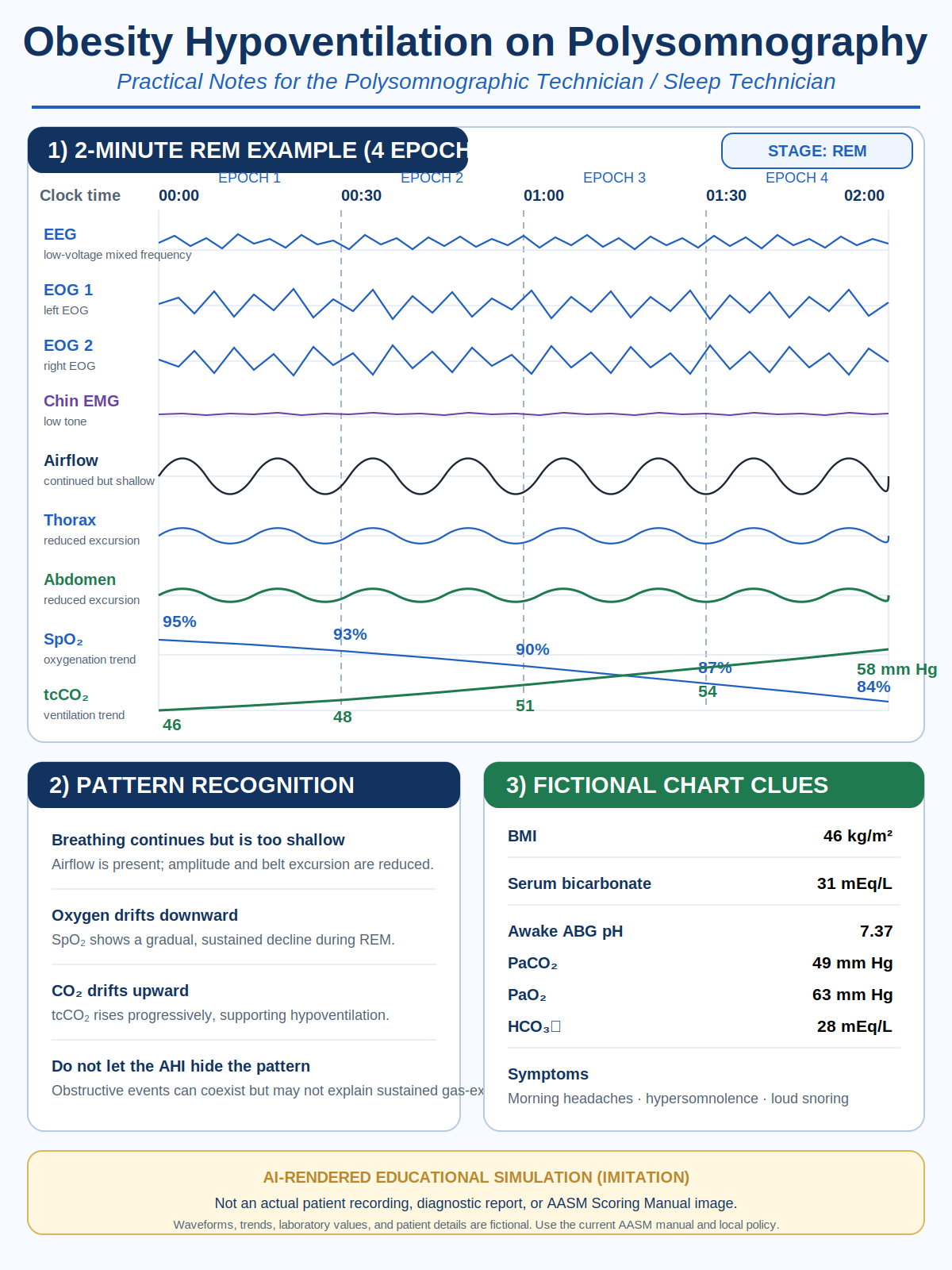
Image disclosure: AI-rendered educational simulation imitating a PSG display. It is not an actual patient recording, diagnostic report, or AASM Scoring Manual image. A two-minute screen can demonstrate pattern recognition, but it cannot independently satisfy a ten-minute adult scoring duration.
3. Adult sleep-hypoventilation scoring anchors
Pathway 1: PCO₂ >55 mm Hg for ≥10 minutes
Pathway 2: PCO₂ rises ≥10 mm Hg from the awake supine baseline to >50 mm Hg for ≥10 minutes
Use PaCO₂ or an accepted surrogate according to the current AASM Scoring Manual and laboratory policy. Validate the CO₂ channel before scoring. A sudden isolated jump without supporting changes should prompt a technical check for sensor contact, drift, calibration, sampling problems, leak, or artifact.
4. Bilevel PAP/NIV, gas exchange, and low-flow oxygen
Ventilation and oxygenation are related—but they are not identical
Bilevel PAP/NIV can improve ventilation by providing pressure support. Increased tidal volume and minute ventilation may improve CO₂ clearance. EPAP also helps maintain upper-airway patency when obstruction is present.
Even when ventilation improves, SpO₂ may remain low. Bilevel PAP/NIV does not automatically correct every cause of impaired oxygen transfer. Persistent hypoxemia may reflect:
- ventilation-perfusion mismatch;
- low functional residual capacity and dependent atelectasis associated with severe obesity;
- residual upper-airway obstruction, leak, or incomplete ventilatory support;
- coexisting pulmonary vascular, cardiac, or lung disease;
- another baseline indication for supplemental oxygen.
Low-flow oxygen does not increase tidal volume, remove retained CO₂, or correct alveolar hypoventilation. In susceptible patients, excessive oxygen may worsen hypercapnia. Continue to evaluate ventilation, CO₂, respiratory effort, leak, stage, position, and the patient—not SpO₂ alone.
Verify signals → control obstruction → optimize ventilation → confirm clinically significant hypoxemia persists → add oxygen only under the physician order and facility protocol → reassess SpO₂ and CO₂ together.
Source basis: Simonds AK. “Indications for Additional Oxygen Treatment,” in the ERS Handbook of Respiratory Sleep Medicine, 2nd ed., pp. 302–303; and Fundamentals of Sleep Technology, 3rd ed., supplemental oxygen and PAP-titration content.
5. Treatment map
| Clinical pattern | General treatment concept | Technologist focus |
|---|---|---|
| Stable OHS with severe OSA | CPAP is commonly the initial therapy. | Control obstruction and verify that oxygenation and CO₂ improve. |
| Predominant hypoventilation, no/milder OSA, or inadequate response to CPAP | Bilevel PAP/NIV may be selected to support ventilation. | Assess EPAP, pressure support, backup strategy when ordered, synchrony, leak, tidal breathing, CO₂, and SpO₂. |
| Persistent hypoxemia after ventilation is optimized | Prescribed low-flow oxygen may be added. | Follow the ordered oxygen protocol and continue CO₂ monitoring. |
| Long-term disease modification | Sustained weight loss through lifestyle treatment, anti-obesity medication, and/or metabolic-bariatric surgery when appropriate. | Do not reduce PAP, NIV, or oxygen without objective reassessment and a new order. |
6. Technologist workflow
- Review the order, chart clues, prescribed oxygen, PAP history, medications, and safety risks.
- Establish and document a valid awake supine CO₂ baseline when required.
- Calibrate, place, and verify airflow, effort, oximetry, CO₂, and PAP/NIV channels.
- Follow stage, position, airflow, effort, oxygenation, CO₂, leak, and patient condition across the night.
- Correct artifact and document interventions objectively.
- Follow the physician order, emergency procedures, and facility titration protocol. Do not independently diagnose or prescribe.
7. Quick RPSGT-style checks
1. Awake supine tcCO₂ is 41 mm Hg. During sleep it rises to 52 mm Hg for 14 minutes. Does this meet an adult scoring pathway?
Yes. The rise is 11 mm Hg, the sleep value is above 50 mm Hg, and the duration is at least 10 minutes.
2. SpO₂ is 84% for six minutes, but no CO₂ channel is available. Is sleep hypoventilation confirmed?
No. Sustained hypoxemia is present, but oximetry alone does not establish hypoventilation.
3. Obstructive events are controlled on bilevel PAP/NIV, but tcCO₂ continues to rise. Is the titration complete?
No. Control of the AHI does not prove adequate ventilation. Verify signals and follow the ordered hypoventilation/NIV protocol.
4. Bilevel PAP/NIV improves tcCO₂, but SpO₂ remains below the prescribed target. What should be considered?
First confirm adequate obstruction control, ventilation, leak, signal validity, stage, and position. When clinically important hypoxemia persists, prescribed low-flow oxygen may be added under the physician order and facility protocol.
5. Oxygen raises SpO₂ from 85% to 92%, while tcCO₂ continues rising. What does that mean?
Oxygenation improved, but ventilation remains inadequate. A better saturation number does not prove that CO₂ clearance has normalized.
6. Which wording is most appropriate for a technologist?
Objectively describe stage, position, airflow, effort, oxygenation, CO₂ trend, signal verification, patient condition, and actions taken. Avoid independently diagnosing OHS or prescribing home therapy.
References and study boundaries
- American Academy of Sleep Medicine. The AASM Manual for the Scoring of Sleep and Associated Events, current version.
- Berry RB, et al. Rules for scoring respiratory events in sleep: update of the 2007 AASM manual. J Clin Sleep Med. 2012;8(5):597–619.
- Mokhlesi B, et al. Evaluation and management of obesity hypoventilation syndrome: an official ATS clinical practice guideline. Am J Respir Crit Care Med. 2019;200(3):e6–e24.
- Masa JF, et al. Obesity hypoventilation syndrome. Eur Respir Rev. 2019;28:180097.
- Simonds AK. Indications for Additional Oxygen Treatment. In: Bonsignore MR, Randerath W, Schiza SE, Simonds AK, eds. ERS Handbook of Respiratory Sleep Medicine. 2nd ed. European Respiratory Society; pp. 302–303.
- Mattice C, Brooks R, Lee-Chiong TL, eds. Fundamentals of Sleep Technology. 3rd ed. AAST/Wolters Kluwer; 2020.
Independent educational resource: Sleep Pathways Guild is not affiliated with, endorsed by, or sponsored by BRPT, AASM, AAST, ATS, or any examination provider. This mini lesson does not reproduce proprietary scoring-manual text and does not replace the current official manual, physician order, device instructions, formal training, clinical supervision, or facility policy.